Tiny larvae were sent by mail from a laboratory in Campinas, in the interior of São Paulo, Brazil, to hospitals in cities such as Natal, Rio de Janeiro, Petrópolis, Belo Horizonte and Porto Alegre.
They were larvae of two species of flies created, fed and sterilized by biologist Patricia Thyssen, from the State University of Campinas (Unicamp), for a very specific medical purpose: to heal wounds that are difficult to heal.
The reason is that these larvae feed on decomposing human tissue.
Thus, when placed on the skin of infected wounds – caused, for example, by diabetes or venous ulcers – the larvae eat the dead tissue and secrete healing substances, reducing the use of antibiotics or even making them unnecessary.
This technique, known as larval therapy, which is still in its infancy in Brazil, has its roots in ancestral, if somewhat repulsive, knowledge: there are historical records that peoples such as the Mayas in Central America and the Australian aborigines were already using larvae to heal wounds thousands of years ago.
The Mayans, for example, bathed tissues in animal blood, left them exposed to the sun to attract flies and then applied them to human wounds, where the larvae proliferated.
The technique was also documented empirically by physicians in medieval Europe, the U.S. Civil War (1861-65) and World War I (1914-18).
Until, in the 20th century, penicillin and the antibiotic revolution made such treatments obsolete.
The problem is that today more and more antibiotics are losing their effectiveness against resistant bacteria, something the World Health Organization (WHO) treats as one of today’s top ten public health threats.
As a result, more healthcare professionals have turned to larvae in recent decades to treat chronic, infected wounds that are resistant to antibiotics and traditional dressings.
In Brazil, researchers want the National Health Surveillance Agency (Anvisa) to validate this type of therapy, as it currently does not classify this type of treatment as a drug or medical therapy.
But it is a treatment that faces many obstacles, and has associated risks, as explained below by BBC News Brazil.
Larvae eating infected tissue
The first clinical study of larval therapy was conducted by the American physician William Baer, based on his experience treating soldiers in France during World War I in 1917.
In a frontline hospital, Baer encountered two patients who, at first glance, appeared to be in a particularly difficult situation: they were soldiers with open wounds in the leg and abdomen, who had spent days in the trenches without treatment, water or food, exposed to unsanitary conditions.
However, Baer noticed that the wounds of the two soldiers were infested with maggots. And that, despite the seemingly bleak picture, the two men arrived at the hospital without fever or signs of septicemia or serious infections.
On the contrary, “when I observed the extent of the wounds, particularly on the thigh, I could not help but marvel at the good condition of the patients,” Baer wrote in his study.
From that episode, the American physician decided to test in the laboratory the effect of the larvae on the wounds, identifying the healing capacity of some of them, although it is important to point out that the lack of sterilization of the larvae used by Baer ended up causing serious secondary infections, such as tetanus, in some of his patients.
Sterilization
More than a century later, today’s larvae therapy is very different, and much more hygienic than that performed by Baer or the Mayan peoples, except that the basic input remains the same: flies.
Actually, very specific flies. Out of hundreds of thousands of fly species, the United Kingdom uses only one – the Lucilia sericata – for medicinal treatment.
It is a species known to breed in garbage and decomposing bodies. And that enables its larvae to treat chronic human wounds, Dr. Yamni Nigam, Professor of Biomedical Sciences at Swansea University (UK), explains to BBC News Brazil.
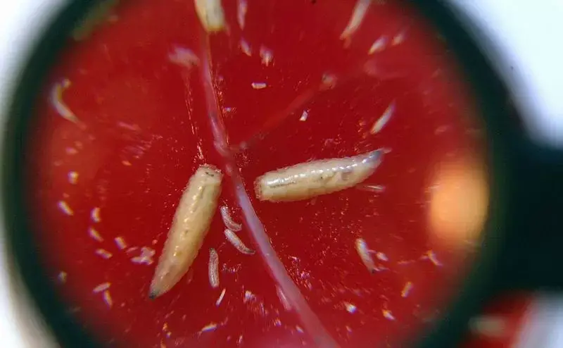
“They feed on these infected and necrotic tissues, clean the wound and stimulate the formation of good skin,” says Nigam.
The primary use is in patients with diabetes, whose wounds, if left untreated, can lead to limb amputation or death.
“These are wounds that just don’t heal, and sometimes the patient doesn’t even realize it, because the nerves (in the injured area) don’t work, there’s a neuropathy. It’s a classic case for the use of larvae,” Nigam explains.
The larva of the Lucilia sericata fly is a non-invasive species, incapable of parasitizing the human body, says the physician. “And she doesn’t eat healthy tissue, so she’s perfect for the job.”
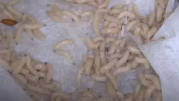
In the UK, treatment is carried out with laboratory-disinfected larvae collected in small, permeable, biological tea bag-like bags.
Under medical supervision, these bags are placed for up to five days over the infected wound and then discarded.
The porosity of the bags allows the larvae to come into direct contact with the wound and, by feeding on this diseased debris, can quadruple in size from 3 millimeters to 12 millimeters.
“The larvae have no teeth: they just secrete a liquid that goes through the sac, and digests and cleans the wound. And then they swallow the liquid again, always inside the pouch,” Nigam continues, citing studies that indicate that the treatment is able to prevent amputations and reduce the need for antibiotics.
Larvae therapy began to be used by some British Public Health Service (NHS) hospitals from the 2000s, at the same time as it was approved by the U.S. drug regulatory agency (FDA).
The British larvae are grown, sterilized and packaged by Welsh company BioMonde, which told BBC News Brazil that it supplies more than 5,000 biological bags to the NHS annually.
The company also has a unit in Germany that exports larvae to European countries.
According to the NHS, in some cases the treatment causes increased local pain, skin irritation or bleeding, and in those cases the larvae must be removed.
“Larvae produce anticoagulants, so we cannot use them in patients at high risk of bleeding,” says Dr. Nigam.
Finally, it is important to emphasize that a treatment of this type should never be performed outside the medical field, and the larvae must be sterilized in a laboratory, warns the Brazilian Patricia Thyssen.
“You should never use a wild larva, because (someone who is not an expert) has no way of knowing if it is a harmless and safe larva species, nor the amount of bacteria that larva can bring,” he clarifies.
But if the risks of the therapy are less than the potential benefits (prevention of amputations and generalized infections, for example), why is the therapy so restricted?
“Disgusting.”
“It’s an underutilized treatment,” Yamni Nigam confirms. “We’ve only used it on very difficult wounds, which are otherwise untreatable. And that’s something we’re trying to change.”
“Why do we leave larvae therapy only as a last resort? Why do we wait for some patients to suffer for years, sometimes trying different types of dressings and ointments, when it would be enough to use larvae for four days?” she questions.
Last year, Nigam and colleagues conducted an opinion poll in the UK, in which only 36% of the 412 respondents said they would agree with using maggots to treat a hypothetical painful wound.
“The main concern is the disgust associated with the therapy,” the survey says.
Other difficulties listed by Nigam are that, unlike traditional medicines and ointments, maggots are not as easily produced and stored, and they often meet resistance among doctors and nurses.
But the superbug breakthrough, according to the physician, has given impetus to new research.
“Bacteria are very intelligent beings. There are few antibiotics left that work against certain diseases. In addition, bacteria settle in wounds and form a wall, what we call bacterial biofilm, something that is very resistant to antibiotics and very difficult to treat,” Nigam explains.
“But we were able to demonstrate, in the laboratory and in patients, that the larvae not only manage to break up this biofilm, but that their liquid also prevents it from forming.”
As this knowledge advances, she says, it may be possible in the future to use the fluid secreted by the larvae to waterproof human prostheses before surgery, for example, to prevent infection.
Nigam believes we need to change the way we view these creatures.
“It seems to me that (larvae therapy) will never become widespread because of the disgust factor, the reluctance,” he says. “But I think the negative perception associated with maggots needs to change. We need to think of them as medicine or medical equipment, not as a repulsive being that we see in the garbage.”